Hip joint is a ball and socket joint that is covered with large muscles of the thighs, lower back, and buttocks. It often takes a significant amount of force to damage a healthy hip joint. Most sports injuries of the hip are a consequence of chronic overuse of the joint and its associated muscles. Any hip injury results in debilitating pain and loss of hip function, and requires a specialist to ascertain early and accurate diagnosis of the injury and initiation of early treatment.
Dr Gautam Tawari is a UK fellowship trained Orthopedic Doctor in Mumbai and the Royal college of surgeons (UK) accredited Orthopedic Sports and Hip arthroscopy surgeon. He uses modern surgical concepts for treatment of labral tears, ligament tears, tendon injuries, osteoarthritis, post-traumatic arthritis, and fractures / trauma around the hip joint.
A sprain is an injury of the ligament and strain is injury of the muscles. Strains and Sprains are common types of injury in overuse conditions, they can be from trauma as well. They are classified as –
Grade I – Mild stretching or microscopic tears accompanied by mild pain. The joint functions normally.
Grade II – Moderate stretching or tears accompanied by pain. The hip may periodically give out while standing or walking.
Grade III – The ligament, muscle, or tendon is completely torn. The hip can no longer bear weight.
Patients often have hip pain and weakness, with activities walking or climbing stairs. Patients can experience muscle spasm, swelling around the hip and bruising. Difficulty or a limp with walking
Though majority of these patients have mild to moderate degree of injury and are commonly seen with repetitive use of the lower body, such as cycling, running, swimming, baseball, and golf.
However, if there is pain and swelling, that doesn’t settle after a few weeks, or if causes severe debilitation, specialist opinion is required for accurate diagnosis and early treatment of the condition.
Inflammation of the tendons or strong tissues around the hip joint is referred to as Hip tendonitis. This inflammation is often due to increased blood supply to the tissue following injury to the tendons. This can also be seen due to degradation of the tendon.
Tendinitis are also part of overuse injuries to the hip. Long-distance runners, swimmers, baseball players, tennis players, and golfers have a higher than average risk of these conditions.
Clinical Presentation includes hip pain, back and leg pain, hip stiffness, hip swelling, Lumps around the hip joint.
Treatment includes use of NSAIDS medications, activity modification and rest. IF the symptoms do not settle down in 3 weeks, injections of PRP can be considered, but a thorough assessment from a specialist is mandatory , who can suggest an appropriate course of management of the problem.
One can lower the risk of recurrent injuries by adequately stretching before exercise and strengthening the muscles around hips, legs, and lower back.
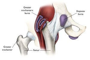
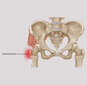
A bursa is a fluid filled sac present around the prominent muscles, bones and tendon. Hip bursa is present on the outer bony prominence of the hip joint. Inflammation of this bursa is called bursitis. Similar to other inflammatory conditions, bursitis is commonly caused by chronic overuse of a joint. Although less likely, bursitis can also occur from acute trauma or infection.
The bursitis pain is often felt in the hip or thigh, outside the hip or thigh, or on one side of the groin. This hip pain and stiffness becomes worse with repetitive motions, hip pain on pressing on the hip or in the groin, Hip Pain when lying on the side or walking up stairs Is common presenting complaints.
The symptoms of bursitis only often last for a few weeks, if the hip is allowed adequate rest. Strengthening of hip, leg, and back muscles help prevent future episodes of bursitis. If bursitis is not settling down, opinion of the specialist should be undertaken for accurate diagnosis and early treatment of the hip pain.
Hip arthroscopy is a surgical procedure that allows an inside view of the hip joint without making a large incision (cut) through the skin and other soft tissues.
Hip Arthroscopy is used to diagnose and treat a wide range of hip problems but requires advanced arthroscopic skills and training to undertake these treatment procedures.
In hip arthroscopy, a small camera, called an arthroscope, is inserted into the hip joint. The camera displays the inside condition of the hip joint on a video monitor. Other surgical instruments (similar to the camera) are inserted in this visual guidance inside the hip joint by making small cuts on the skin.
These instruments are used to treat the underlying condition causing the hip pain. This results in less pain, less joint stiffness, and shortens the time it takes to recover and return to activities and sports.
Hip arthroscopy is not as common as knee or shoulder arthroscopy and fairly more complex due to the reduced space in the hip joint. This is recommended to be done by specialists only.
Hip arthroscopy is used to relieve hip pain arising from damage the labrum, articular cartilage, or other soft tissues surrounding the joint.
Patients often have one of the following diagnosis that requires hip arthroscopy –
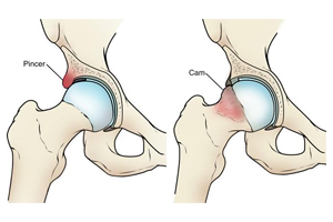
- Femoroacetabular impingement (FAI) – A condition where extra bone develops along the acetabulum (pincer impingement) or on the femoral head (cam impingement). These bone overgrowth damages the tissues of the hip during movement.
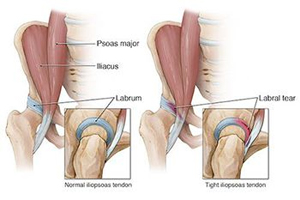
- Hip Dysplasia - A condition where the hip socket is abnormally shallow. This puts more stress on the labrum (outer cushion ) that keeps the femoral head within the socket, and makes the labrum susceptible to tearing and damage.
- Snapping hip syndromes – This condition is caused by a tendon rubbing across the outside of the joint. There is snapping or popping noise present with certain movements of the hip joint. It is often harmless but in some cases, however, the tendon is damaged from the repeated rubbing.
- Hip Synovitis – This is a painful condition resulting from the tissues surrounding the hip joint to become inflamed.
- Loose bodies – These are fragments of bone or cartilage that become loose and move around within the joint.
- Hip joint infection
Post op Recovery
After surgery, the patient stays in the hospital for the next 6 to 8 hrs before being discharged home. It is expect that the patient uses crutches, or a walker, for some period of time.
Patients return to full, unrestricted activities after arthroscopy. the recovery however depends upon the type of damage that was present in your hip and the complexity of the procedure undertaken to address the problem.