The Orthopedic knee joint is the commonest joint to be injured with playing sports, the bones of the knee are covered with cartilage to allow smooth movement. There is medial & lateral meniscus (cushions) – that function as shock absorbers. It has two internal cruciate ligaments (ACL & PCL) and two external collateral ligament (MCL & LCL) that provide stability.
Severe injuries to the knee joint requires a review from a Knee specialist to ascertain early and accurate diagnosis of the injury and initiation of early treatment. Dr Gautam Tawari is a UK fellowship trained and a Royal college of surgeons accredited Orthopedic Sports and Knee arthroscopy surgeon in Mumbai. He undertakes arthroscopic ACL/PCL surgery, Meniscal repairs, Knee Cartilage surgery and Stem cell treatment in the knee joint.
He follows an advanced rehabilitation protocol to ascertain his patients get back to sports quicker and more confidently.

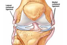
The collateral ligaments connect the femur to tibia. It control the sideways motion of the knee and braces against unusual movement. The knee has 2 collateral ligaments – medial or “inside” collateral ligament and the lateral or “outside” collateral ligament (LCL). Injury to ligaments are considered as “sprains” and are graded.
Grade 1 Sprain
– The ligament has been slightly stretched, but is still able to help keep the knee joint stable.
Grade 2 Sprain
– The ligament stretches to a point where it becomes loose. This is often referred to as a partial tear.
Grade 3 Sprain
– The ligament is split into two pieces. This is often referred as complete tear.
Cause
Injuries are often contact injuries, but not always. Medial collateral ligament tears occur as a result of a direct blow to the outside of the knee. Blows to the inside of the knee tears the lateral collateral ligament. The MCL is injured more often than the LCL.
Presentation
- Pain at the sides of your knee. If there is an MCL injury, the pain is on the inside of the knee; an LCL injury may cause pain on the outside of the knee.
- Swelling over the site of the injury.
- Instability — the feeling that your knee is giving way.
Treatment
Initial treatment includes icing, knee brace, activity modification, physiotherapy and painkillers.
Surgery is advocated on failure of conservative treatment, location of the tear in the ligament or associated injuries. Chronic instability require ligament reconstruction.

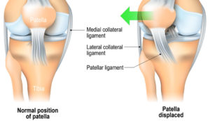
Kneecap (patella) rests in a groove of the thighbone (femur). When the knee bends and straightens, the patella moves straight up and down within the groove.
Causes
- Direct blow or a fall onto the knee, common in high-impact sports, such as football.
- A shallow or uneven groove in the femur can make dislocation more likely.
- Loose ligaments, making joints extremely flexible and more prone to patellar dislocation. This id common in girls and affects both the knees.
- Conditions causing muscle imbalance like Cerebral palsy, Down syndrome
Presentation
- Pain, popping sound when the patella dislocates
- Feeling of kneecap shifting or sliding out of the groove, knee buckling or giving way
- Swelling, deformation of the knee
- Apprehension or fear when running or changing direction.
Treatment
Dislocation often damages knee tissue and the patella remains unstable. Exercises, such as cycling, can help strengthen quadriceps muscles in the thigh and prevent future patellar dislocations.
Multiple dislocation or instability warrants surgery. Surgical treatment involves arthroscopic (keyhole) reconstruction of the ligaments that hold the patella in place. Presence of bone deformity require more complex surgical treatment.